


February 2, 2022 -- The cellular barrier that protects the brain also has the unfortunate effect of preventing medical treatments from reaching areas of the brain where they are needed. This presents a major obstacle in developing new drugs to treat brain diseases. Now, in a study published in Nature Protocols, researchers describe an approach in mice to precisely open a gate in the barrier and deliver therapeutic agents.
The blood-brain barrier is a protection mechanism in vertebrates to prevent pathogens and toxins from harming the brain. While it is not entirely effective -- small molecules like ethanol can still reach brain cells -- it does block many druglike compounds. However, this barrier creates great difficulty for scientists trying to develop new treatments for diseases, such as brain tumors, Alzheimer's disease, or Parkinson's disease.
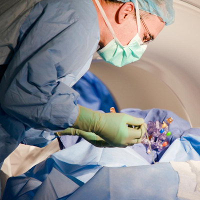
One way to open the barrier is by injecting the sugar mannitol through a catheter that runs along the carotid artery and into the brain. A high concentration of mannitol causes the cells that make the barrier to shrink and reveal gaps that could allow large molecules and cells through.
Although this approach has been used for decades in lab animals, its results are highly variable and inconsistent. The dynamics of brain blood flow can be different between individuals, and an injection rate that is too high could break the barrier rather than creating a temporary gate.
"Researchers had no way of knowing if the drug made it where it was supposed to go at the time of the experiment and some researchers found the procedure unsafe in animals," said Dr. Miroslaw Janowski, PhD, associate professor at the University of Maryland School of Medicine who co-led the study, in a statement. (Nat Protoc, December 13, 2021, Vol. 17, pp. 76-94.)
Fine-tuning the opening
To improve the safety and precision of this approach, Janowski and colleagues developed a technique using magnetic resonance imaging (MRI) scans to fine-tune the injection. Inserting a catheter through the carotid artery in anesthetized mice, the team injected a compound containing the element gadolinium, which provides a strong signal on MRI scans. This allowed researchers to optimize the injection rate and thereby control the opening in the blood-brain barrier when the mannitol was injected.
"Arterial procedures are typically guided using X-ray imaging methods, and that only lets you see the map of the highways in the brain, but these maps are not detailed enough if you needed to go beyond that to the backroads to reach the forest of the brain cells," Janowski said. "We believe that by adding MRI, we closed the gap, both in animal studies and in the future in the clinical trials, by providing the necessary level of precision for opening the gate to the brain for the drugs."
The team deliberately chose to test this procedure in mice, despite it being more technically challenging than in rats or large animals, because mice are so widely used to study neurodegenerative disorders. Opening the blood-brain barrier in the animals increased the dosage into the brain of large therapeutics, such as monoclonal antibodies.
The published paper describes the technique in detail so that other laboratories can use it to investigate a variety of treatments and diseases.
Benefits for patients
The researchers previously applied this MRI-guided approach in a patient with a brain tumor to deliver a chemotherapy drug directly into the brain. In humans, this is a minimally invasive procedure, as the catheter can be inserted via the femoral artery by the groin. However, more research would be needed to show that the MRI guide is safe and reliable enough for regular clinical use.
"Scores of interventional radiologists worldwide navigate various sophisticated devices in arteries for standard plumbing tasks in the human brain, like aneurysms or strokes. If there is a leak, they fix it. If there is a clog, they pump it out. This route could be also used to deliver drugs, but the technique had not been perfected in mice and other laboratory animals," said senior author Dr. Piotr Walczak, PhD, a professor at the University of Maryland School of Medicine.
"Since most preclinical research starts with mice, it was essential to show how to do this procedure properly in these animals so scientists across the globe will be able to use it for their drugs to get them to the brain. Some of these drugs will ultimately land in patients as treatments benefiting society."
Do you have a unique perspective on your research related to preclinical research? Contact the editor today to learn more.
---

Copyright © 2022 scienceboard.net
Member Rewards
Earn points for contributing to market research. Redeem your points for merchandise, travel, or even to help your favorite charity.

Research Topics
Interact with an engaged, global community of your peers who come together to discuss their work and opportunities.
Conferences
Connect


