


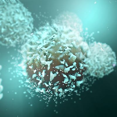
September 8, 2022 -- German researchers have used cryo-electron microscopy (cryo-EM) to visualize biomolecules at the atomic level, combining cryo-EM with a method used in materials research to observe protein building blocks in their natural environment in a snap-frozen state.
The team from Forschungszentrum Jülich and Heinrich Heine University Düsseldorf have combined cryo-EM with integrated differential phase contrast scanning transmission electron microscopy (iDPC-STEM).
In a study published on September 5 in the journal Nature Methods, the scientists applied iDPC-STEM to two cryo-EM test specimens: keyhole limpet hemocyanin (KLH) and tobacco mosaic virus (TMV). The micrographs showed complete contrast transfer to high resolution and enabled the cryo-EM structure determination for KLH at 6.5 Å and 3.5 Å for TMV using single-particle reconstruction methods.
The findings demonstrate that STEM imaging in general and the iDPC-STEM approach in particular can be applied to vitrified single-particle specimens to determine near-atomic resolution cryo-EM structures of biological macromolecules. This expands the possibilities for structural analysis, especially for heterogeneous, nonuniform samples or single particles when averaging capabilities are limited, the researchers noted.

Copyright © 2022 scienceboard.net
Member Rewards
Earn points for contributing to market research. Redeem your points for merchandise, travel, or even to help your favorite charity.

Research Topics
Interact with an engaged, global community of your peers who come together to discuss their work and opportunities.
Conferences
Connect


