


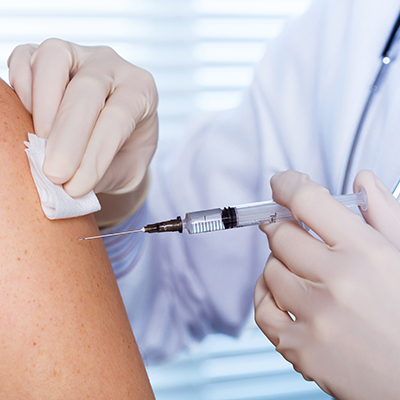
December 7, 2022 -- Duke University and Albert Einstein College of Medicine researchers have designed a small fluorescent protein that absorbs and emits wavelengths of near-infrared (NIR) light that penetrate deep into living tissue. Their technique, outlined December 1 in the journal Nature Methods, captures detailed high-resolution biomedical images.
Visible light is quickly absorbed and scattered, preventing researchers from seeing deeper than a millimeter within tissue. The researchers focused on the 700 nm to 1300 nm window of NIR light, where tissue is most transparent.
The team used a directed molecular evolution process to engineer proteins, using bacterial photoreceptors as a structural basis. They studied the biomolecule biliverdin, abundant in mammalian tissues, to determine how the photoreceptors could best attach to it. Substitutions of key molecule parts connecting to the biliverdin increased the electron binding necessary to obtain red-shifted fluorescence. Finally, random mutagenesis, followed by high-throughput screenings, enabled the proteins to evolve and increase their brightness.
The brightest found protein, miRFP718nano, is produced in cells and emits light just outside the visible range. When NIR light first hits these proteins, they emit light in the first 700 nm to 900 nm zone. But as they decay, they begin emitting light in the second 900 nm to 1300 nm zone. Here, light penetrates tissue two times deeper, background fluorescence is dimmed, and image resolution clearer, allowing more detailed images of complex structures than standard NIR zone-one imaging proteins.
A short-wavelength infrared imaging technique tested miRFP718nano's efficacy. After introducing engineered miRFP718nano into mice, the team captured images of digestive tract microbes, visualized mammary gland cells, and tracked liver inflammation. The technique may eventually help visualize the brain and track cancer cell movement.
"This is an exciting new front of our decade-long collaboration because we can use the imaging tools to guide the protein engineering decisions, and we can use the advanced protein engineering to improve the imaging capabilities," co-author Junjie Yao, PhD, assistant professor of biomedical engineering at Duke, said in a statement.


Copyright © 2022 scienceboard.net
Member Rewards
Earn points for contributing to market research. Redeem your points for merchandise, travel, or even to help your favorite charity.

Research Topics
Interact with an engaged, global community of your peers who come together to discuss their work and opportunities.
Conferences
Connect


