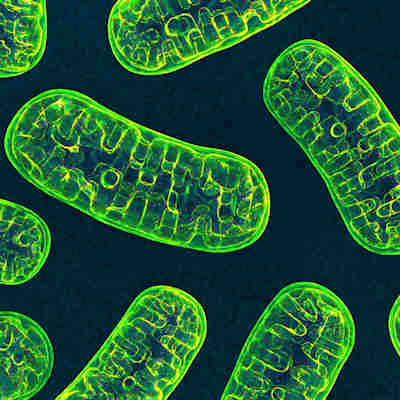
August 8, 2022 -- Researchers from the National Institutes of Health (NIH) have used cryo-electron microscopy (cryo-EM) and other techniques to create a 3D structure of the twinkle protein, the final piece of the human minimal mitochondrial replisome to be structurally characterized.
The protein, the helicase in the mitochondrial DNA replisome, helps the body use energy converted from food. Mutations to twinkle can lead to mitochondrial diseases but, working from models, researchers and clinicians were previously unable to determine the mechanistic links.
By creating 3D structures of human twinkle W315L, the researchers have provided the means to show the locations of mutations and to develop targeted therapies for conditions such as progressive external ophthalmoplegia, Perrault syndrome, and hepatocerebral mitochondrial DNA depletion syndrome.
The researchers from NIH's National Institute of Environmental Health Sciences (NIEHS) described their work in an article published in the journal Proceedings of the National Academy of Sciences. The paper describes how the team used cryo-EM to characterize the oligomeric assemblies of human full-length twinkle W315L, define its multimeric interface, and map clinical variants associated with the protein in inherited mitochondrial disease. W315L is a clinical mutation known to cause progressive external ophthalmoplegia.
Using cryo-EM, lead author Amanda Riccio, PhD, researcher in the NIEHS Mitochondrial DNA Replication Group, and her collaborators observed thousands of protein particles appearing in different orientations and revealed a final structure with a multiprotein circular arrangement. The team used crosslinking-mass spectrometry to verify the structure and molecular dynamics simulations to deliver insights into the dynamic movement and molecular consequences of the W315L clinical variant.
The work revealed that clinical mutations can change the shape of the twinkle pieces and prevent them from fitting together and performing their intended function. Riccio and her collaborators mapped 25 disease-causing mutations, showing that many of the genetic changes map right at the junction of two protein subunits. The finding suggests mutations in this region would weaken how the subunits interact and make the helicase unable to function.
"What is so beautiful about Dr. Riccio and the team's work is that the structure allows you to see so many of these disease mutations assembled in one place," Matthew Longley, PhD, an author and NIEHS researcher, said in a statement. "It is very unusual to see one paper that explains so many clinical mutations. Thanks to this work, we are one step closer to having information that can be used to develop treatments for these debilitating diseases."
The researchers have made the twinkle structure and all the coordinates freely available in the open data Protein Data Bank.
Copyright © 2022 scienceboard.net


