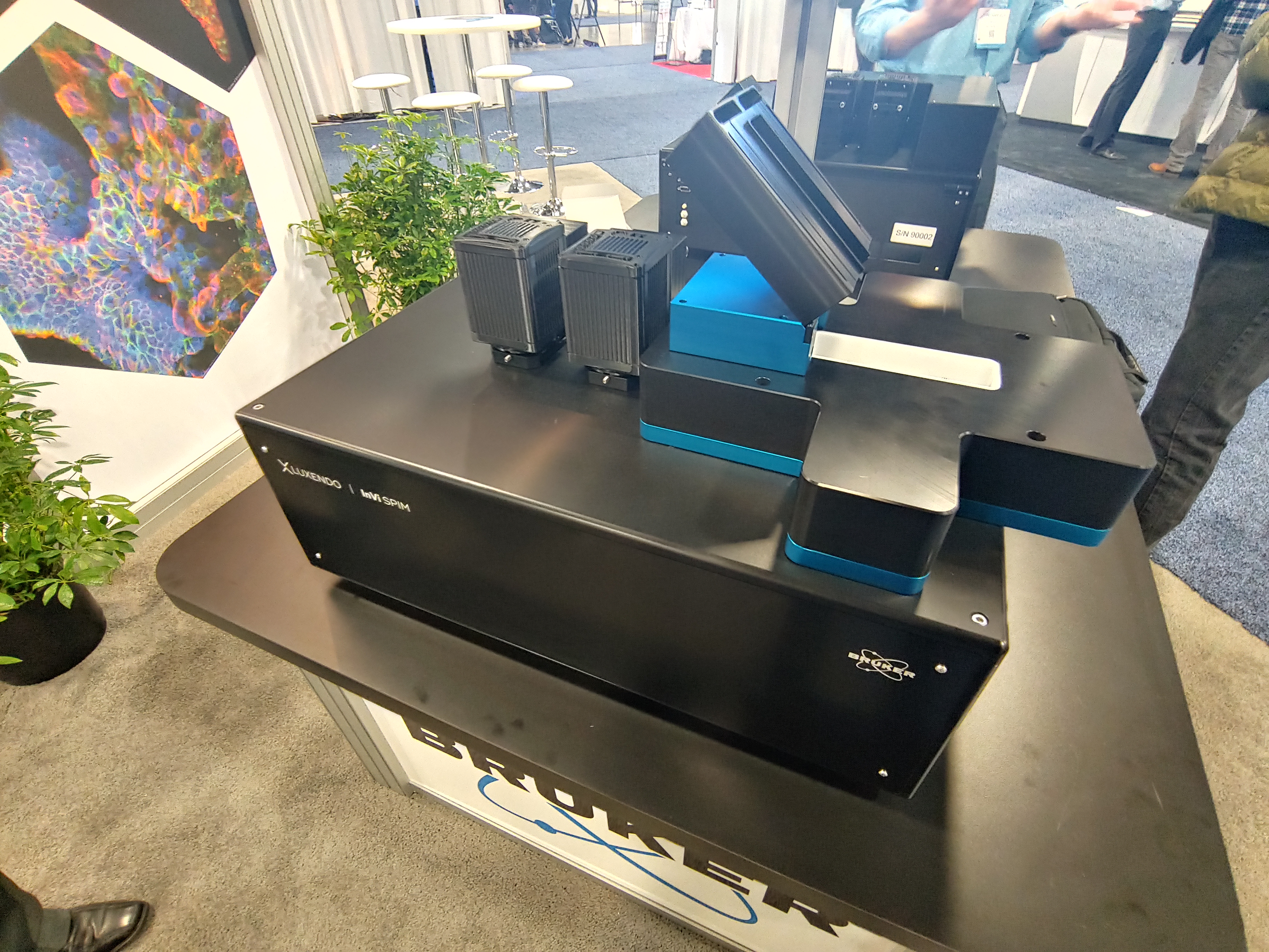
December 11, 2019 -- On Monday, December 10, at the American Society for Cell Biology (ASCB) and European Molecular Biology Organization (EMBO) meeting, Bruker introduced a brand new product which brings increased options to scientists looking for live-cell 3D imaging.

The Luxendo TruLive3D Imager light-sheet imaging system. This dual-sided illumination product allows scientists to capture three-dimensional images of samples with deep penetration of live samples (such as embryos) over time. It leverages the general benefits of single-plane illumination microscopy (SPIM) to enable rapid 3D imaging with minimal light exposure, confocal resolution and excellent contrast in 3D.
"Life happens in 3D," added Dr. Lars Hufnagel, Vice President and General Manager of Bruker's Luxendo business. "Our new TruLive3D Imager now enables imaging biology in its native environment without compromising resolution and imaging speed or worrying about photo-toxicity."
This new system is particularly gentle on samples, providing controlled incubation conditions that enables imaging over multiple days. The large sample chamber (75 mm in length) can accommodate up to 100 samples into the chamber trough and is ideal for multi-position imaging of small embryos (Zebrafish, Drosophila, and mouse) or 3D spheroids.
Other benefits of the system include minimized toxicity and photobleaching effects. Luxendo's new TruLive3D Dish series work seamlessly with the system. The sterile, dual-well dishes are customizable and enable researchers to conduct multiple experiments at once or add controls into their protocols. This product is back by two strong brands and is backed by a breadth of knowledge and expertise in high-performance light sheet microscopy.
Copyright © 2019 scienceboard.net


