


October 31, 2022 -- Weill Cornell Medicine researchers have developed a new computational method to map the architecture of human tissues in unprecedented detail, with the potential to accelerate studies on organ-scale cellular interactions and enable powerful new diagnostic strategies for an array of diseases.
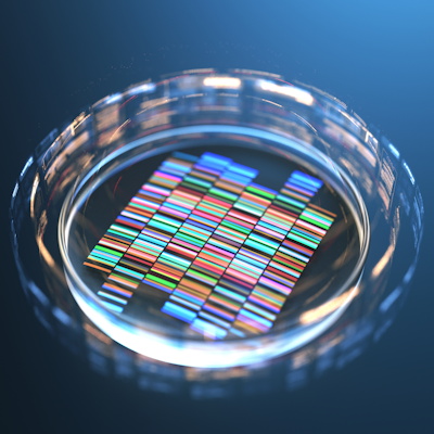
While manually combining single-cell data with maps of tissue structure is a slow and complex process, researchers developed a computational strategy -- called unsupervised discovery of tissue architecture with graphs (UTAG) -- using a combination of single-cell gene expression profiles and cells' locations to define structural regions within tissue.
"We demonstrate that our approach is able to discover organ-specific microanatomical domains in the human lung with high accuracy in comparison with manual annotations and outperforms other methods. Furthermore, UTAG can be employed across various physiological states, such as infectious disease and cancer, and its results can reveal the high-level organization of tissues at the whole-organ scale," the authors wrote in an October 31 study published in Nature Methods.
Using the new method, researchers generated detailed maps of several types of tissues while identifying and quantifying new aspects of microanatomy -- the patterns that emerge at small scale when cells interact and determine the function of tissues. Working with Scott Randell, PhD, a professor at the University of North Carolina School of Medicine's Marsico Lung Institute who studies lung disease, they demonstrated that the computational technique could draw fine shades of distinction between different disease states in a tissue.
Co-senior author André Rendeiro, PhD, a postdoctoral fellow at Weill Cornell Medicine during the study and currently a principal investigator at the Research Center for Molecular Medicine of the Austrian Academy of Sciences in Vienna, Austria, said in a statement that severe COVID-19 is an example in which "there are a lot of immune cells that move into the neighborhood, and there's really dramatic change in the lung tissue."
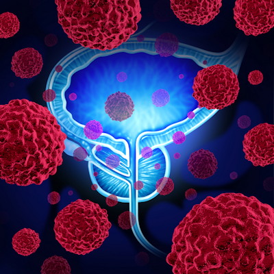
Although cancer and other chronic diseases often cause major changes in tissue structure, the scientists contend that detailed microanatomy could also help in diagnosing and treating more acute conditions.
Going forward, the researchers are applying their new technique to a wide range of tissues to understand how changes in tissue organization underlie its function in healthy state and dysfunction in disease.
"It's crucial for researchers to learn more about the details of tissue structure; fundamental changes in the relationships between cells within a tissue drive both healthy and diseased organ function," said senior author Olivier Elemento, PhD, director of the Englander Institute for Precision Medicine and a professor of physiology and biophysics and of computational genomics in computational biomedicine at Weill Cornell Medicine.
Copyright © 2022 scienceboard.net
Member Rewards
Earn points for contributing to market research. Redeem your points for merchandise, travel, or even to help your favorite charity.

Research Topics
Interact with an engaged, global community of your peers who come together to discuss their work and opportunities.
Conferences
Connect


