


June 30, 2022 -- Researchers at the Massachusetts Institute of Technology’s (MIT) Singapore site have developed a new way to detect adventitious microbial contamination in mesenchymal stromal cell (MSC) cultures. By using machine learning to predict if a culture is contaminated in near real-time, the approach could enable testing to take place during the production of cell therapy products.
Currently, cell therapy manufacturers perform end-point testing. The researchers see the use of the anomaly detection aided, label-free ultraviolet spectroscopy method to detect contamination reducing the time and manpower needed to find problems.
MSCs' ability to self-regenerate, differentiate into cell lineages, and play a role in immunomodulation has attracted researchers who have identified the cells as the basis for potentially effective treatments of a range of diseases. The cell therapy production process needs monitoring to ensure product quality and safety, but the incubation period of established tests is too long to support continuous oversight.
Recognizing the limitations of existing tests, researchers at the Singapore-MIT Alliance for Research and Technology (SMART) aimed to develop a method suitable for integrating with at-line technologies for continuous monitoring.
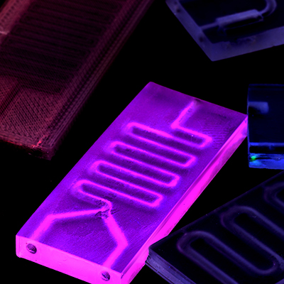
The team developed the machine learning model by collecting sterile cell culture media samples from a range of MSC cultures. Having collected samples from different conditions, the researchers spiked some of them with different bacteria strains at a range of concentrations.
Using ultraviolet-visible spectrometry, the SMART team obtained the absorbance spectra of the sterile, unspiked and bacteria-spiked samples. The researchers then used the spectra to train a machine learning model. In an abstract published in the journal Cytotherapy, the team showed the model achieved 96% accuracy when tested on four sterile and 12 bacteria-inoculated media spectra.
The model achieved that level of accuracy when detecting bacteria at a concentration of 1,000 colony forming units (CFU) per milliliter. The researchers now plan to use bacteria-inoculated samples of lower CFU to improve the sensitivity of the model, as well as work to integrate it and the sampling platform with bioreactors to enable automated periodic sampling and real-time detection.
"Our increased adoption of machine learning in microbial anomaly detection has enabled us to develop a unique test which quickly performs in-process contamination monitoring, marking a huge step forward in further streamlining the cell therapy manufacturing process," said Yie Hou Lee, scientific director of SMART CAMP, in a statement.
The publication of the abstract comes weeks after the release of another SMART paper on the detection of microbial contamination in cell therapy production. In that project, details of which were published on April 13 in the journal Molecular Therapy: Methods & Clinical Development. The researchers used the ratio of nicotinic acid to nicotinamide as a biomarker for assessing sterility.


Copyright © 2022 scienceboard.net
Member Rewards
Earn points for contributing to market research. Redeem your points for merchandise, travel, or even to help your favorite charity.

Research Topics
Interact with an engaged, global community of your peers who come together to discuss their work and opportunities.
Conferences
Connect


