
March 2, 2022 -- Mapping out the intricate network of blood vessels in tissues can help researchers to better understand the changes in diseases like dementia or cancer. However, techniques to image the vasculature are individually limited and difficult to combine. Now, a new easy-to-use system, described in a paper published February 10 in the journal Nature Methods, can combine multiple imaging techniques to provide more detailed information at a range of scales.
The role of blood vessels in bringing oxygen and nutrients to every organ and tissue makes them critically important to understand in both health and disease. The capillaries that reach deep into the brain are key defenses against toxins. Meanwhile, tumors can prompt the growth of new blood vessels to nourish the cancerous cells and maintain their rapid spread.
Traditionally, tracing how blood flows through tissues is challenging. Blood vessels vary enormously in size, from centimeter-wide major arteries down to the fine capillaries that are a thousand times smaller. Standard imaging techniques like computer tomography (CT), magnetic resonance imaging (MRI), and optical microscopy each have their strengths at different scales of resolution. Each scan can be used to answer specific questions about the tissue sample being studied. Getting the full picture, however, requires observing blood vessels at all levels.
Combining scans
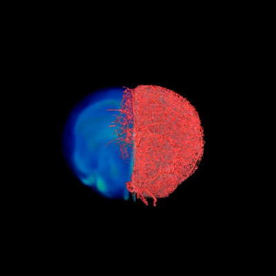
A still from the visual demonstration of the result of the VascuViz imaging pipeline. Image courtesy of Johns Hopkins Medicine.
A combination of these imaging techniques could provide a much more detailed impression of the vasculature of a tissue sample, but this is difficult to achieve. The chemicals added to tissue to provide a signal, known as contrast agents, are specific to each imaging method and generally incompatible with one another. Traditional CT vascular contrast agents tend to be insoluble in water and invisible on MRI, while fluorescent labels used for optical imaging do not contain the chemical groups necessary to be visible in MRI or CT. Advanced methods that can combine these different scans tend to require complex preparation that can limit their widespread adoption.
“Usually, if you want to gather data on blood vessels in a given tissue and combine it with all of its surrounding context like the structure and the types of cells growing there, you have to re-label the tissue several times, acquire multiple images and piece together the complementary information,” commented Arvind Pathak, PhD, professor of radiology, biomedical and electrical engineering and member of the Sidney Kimmel Comprehensive Cancer at the Johns Hopkins University School of Medicine, in a statement.
“This can be an expensive and time-consuming process that risks destroying the tissue’s architecture, precluding our ability to use the combined information in novel ways.”
Pathak and his colleagues have developed a new easy-to-use method, dubbed VascuViz, for combining CT, MRI, and optical microscopy on the same sample. The team tested combinations of contrast agents to find a pairing that could work in parallel. The search found that the CT agent BriteVu and the MRI agent Galbumin-Rhodamin (GalRh) could be combined together. As GalRh is fluorescently labeled, it also provides a signal for optical microscopy. These agents can be bought commercially, making them affordable and readily available.
Creating a 3D map
The team tested VascuViz in a variety of mouse tissues, as well as a human breast cancer model. They could then combine the resulting images from the scans to create 3D visualizations of the vasculature. The technique also worked in tissue sections that were frozen or embedded with paraffin, which are standard treatments for samples in histopathology. This means that the VascuViz system could be easily integrated into existing workflows for tissue analysis.
The method is not limited by the size of the tissue being studied and could be used for whole organs or even a whole mouse. A scan of a mouse brain produced a 3D map that could visualize the whole brain down to single capillaries. This high level of resolution allowed the team to simulate the blood flow in individual brain regions.
“Now, rather than using an approximation, we can more precisely estimate features like blood flow in actual blood vessels and combine it with complementary information, such as cell density,” said Akanksha Bhargava, PhD, a postdoctoral fellow in Pathak’s laboratory.
In the future, the authors suggest, this approach could enable researchers to visualize the response to antiangiogenic cancer therapies, which stop blood vessels from providing nutrients to tumors.
Do you have a unique perspective on your research related to neuroscience or preclinical reserach? Contact the editor today to learn more.
---
Copyright © 2022 scienceboard.net