


November 11, 2020 -- A new digital microfluidic technique allows researchers to connect physical cell properties with the molecular makeup of individual cells. This new approach, published in Nature Communications on November 11, will enable a deeper study of stem cells and other rare cell types for therapeutic development.
Omics analysis has transformed research in the life sciences by enabling the assessment of the genomes, transcriptomes, or proteomes of individual cells. Microfluidic-based technologies that drive this analysis rely on partitioning cells into droplets, microchannels, or microwells where they can be analyzed by any number of omics techniques. While this technology has been transformative, the methodology lacks the capacity to correlate single-cell information with phenotypes of the cells.
To connect single-cell sequencing data to cellular immunofluorescence-based phenotypes, researchers from the University of Toronto developed a new technology called digital microfluidic isolation of single cells for -omics (DISCO). The technology combines digital microfluidics, laser cell lysis, and artificial intelligence-driven image processing to collect the contents of single cells from a heterogeneous population.
Digital microfluidic techniques allow for automated culture, fixation, staining, and image-based analysis of adherent cells in situ. Laser cell lysis is a technique in which a focused, high-energy laser is used to lyse cells. Together, these techniques ablate single-cell contents into capillaries for electrophoretic analysis. Following ablation, single-cell genomes and transcriptomes are determined by next-generation sequencing, and proteomics by nanoflow liquid chromatography and tandem mass spectrometry.
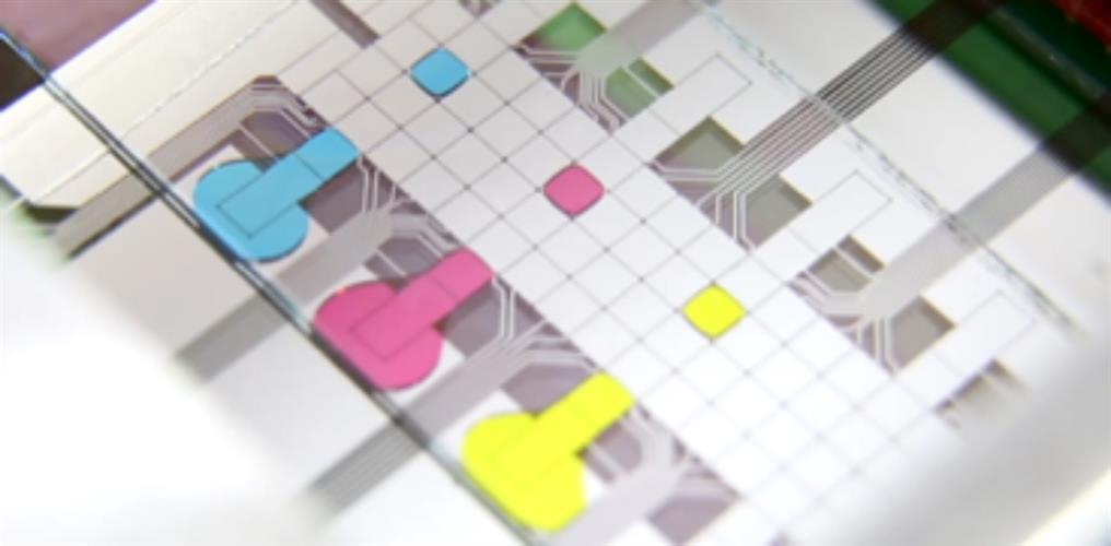
A microfluidic chip with droplets of liquid dyed in different colors for easier visualization. Each droplet can ferry material from individual cells for single cell omics analyses. Image courtesy of the Wheeler lab, University of Toronto.
"We give the user the power to take beautiful fluorescence microscopy images to learn everything that can be learned about cells growing in situ and then connect that information with the cell's genome, transcriptome and proteome," said lead author Aaron Wheeler, PhD, a professor of chemistry and biomedical engineering in the Donnelly Centre for Cellular and Biomolecular Research at the University of Toronto, in a statement.
DISCO is composed of a microscope fitted with a high-frequency laser and a microfluidic chip for the collection of cellular material. Cells and reagents are delivered by passive dispensing, in which droplets spontaneously form virtual microwells containing anywhere from 100 to 300 adherent cells. The user can choose any cell or combination of cells of interest.
After selection and dispensing, the laser is used to rupture the droplet and dispense cell contents into a solution onto the microfluidic chip where it can be retrieved for molecular sequencing.
"Our platform focuses on the metadata that you lose when you do single-cell suspension, things like cell position, what were its morphological properties, who were its neighbors?" said author Erica Scott, PhD, postdoctoral fellow at the University of Toronto. "Those are all the things that we can capture before we do the single-cell sequencing. To our knowledge, this is the only platform that can take cells in culture and do this kind of thing."
The researchers conducted proof-of-concept experiments to demonstrate DISCO's ability to faithfully relate omics data to individual cells cultured as monocultures and side by side. For the experiments they used human glioblastoma (U87) and murine melanoma (B16) cell lines.
In monocultures of cancer cells, they found that there were subtle differences in the copy number variants between cells, validating DISCO's sensitivity at evaluating single-cell genomics. In addition, cocultures revealed that cell-to-cell interactions impact transcriptional signatures of individual cells when human cells are transplanted into foreign cell types (in this case mouse cells), providing insight into cellular relationships.
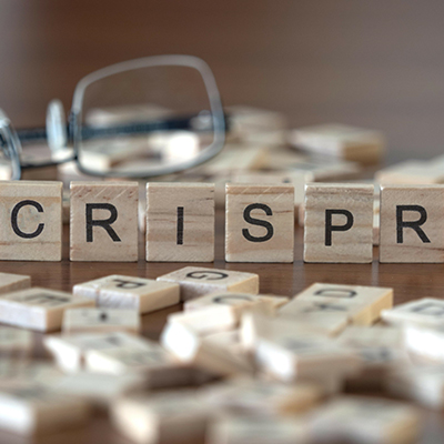
To explore the correlation between phenotype and genotype after genetic modification by CRISPR, the researchers used DISCO to select and lyse single wildtype and knockout cells. Of the eight knockout cells analyzed, four had large deletions, two had medium-size deletions, and two had small deletions.
They also found that CD47 intensity by immunofluorescent labeling was decreased in the knockout cells with deletions compared to the wildtype, indicating that DISCO is capable of associating genomic composition with phenotypic assessments.
According to the authors, the findings can help researchers gain a better understanding of healthy and diseased human tissue, such as tumors, by growing cells in mice to study them in a whole-body environment. The researchers are also working to adapt DISCO to analyze tissue slices. The ultimate goal is to study rare cell types whose regenerative potential is regulated by their immediate environment to advance new therapies.
Do you have a unique perspective on your research related to microscopy or cell biology? Contact the editor today to learn more.
---

Copyright © 2020 scienceboard.net
Member Rewards
Earn points for contributing to market research. Redeem your points for merchandise, travel, or even to help your favorite charity.

Research Topics
Interact with an engaged, global community of your peers who come together to discuss their work and opportunities.
Conferences
Connect


