January 31, 2020 -- Molecular and computational analysis of immune responses in the central nervous system (CNS) reveals that the brain's immune system may prevent blood immune cells from entering the site of a lesion after injury. The research, published online in Science Advances on January 15, may offer new avenues to treat certain neurological diseases such as multiple sclerosis, Alzheimer's disease, and spinal cord injury.
Injury to the central nervous system can activate two types of immune cells, microglia and CNS-infiltrating macrophages. Microglia is a type of macrophage that accounts for 10% to 15% of all cells found in the brain; they are the first form of immune defense in the central nervous system. Meanwhile, CNS-infiltrating macrophages originate peripherally from bone marrow.
However, due to the similarities of these cells, it has been difficult to classify their specific roles in CNS injuries. So, researchers from the University of Alberta, McGill University, and the University of Calgary sought to characterize the activation profile of microglia with fate mapping strategies using reporters and biomarkers.
In this study, microglia and macrophages were labeled with Cre recombinase under the CX3CR1 promoter crossed with the tdTom reporter line. Measuring expression changes of microglia and macrophages after injury, the researchers induced focal demyelination, damage to the myelin sheath, in the spinal cord by injecting lysophosphatidylcholine (LPC). Flow cytometry and immunohistochemistry revealed that the cells were recruited to the site following injury.
To further distinguish between microglia and CNS-infiltrating macrophages, expression of CD45, which is high in infiltrating macrophages, and CX3CR1, which is enriched in microglia, were measured before and after injury. They found that following injury, while CD45 expression was high in both microglia and macrophages, there was a trend of greater CD45 intensity in tdTOM+ cells (microglia) and CX3CR1 levels were downregulated, indicating activation of microglia.
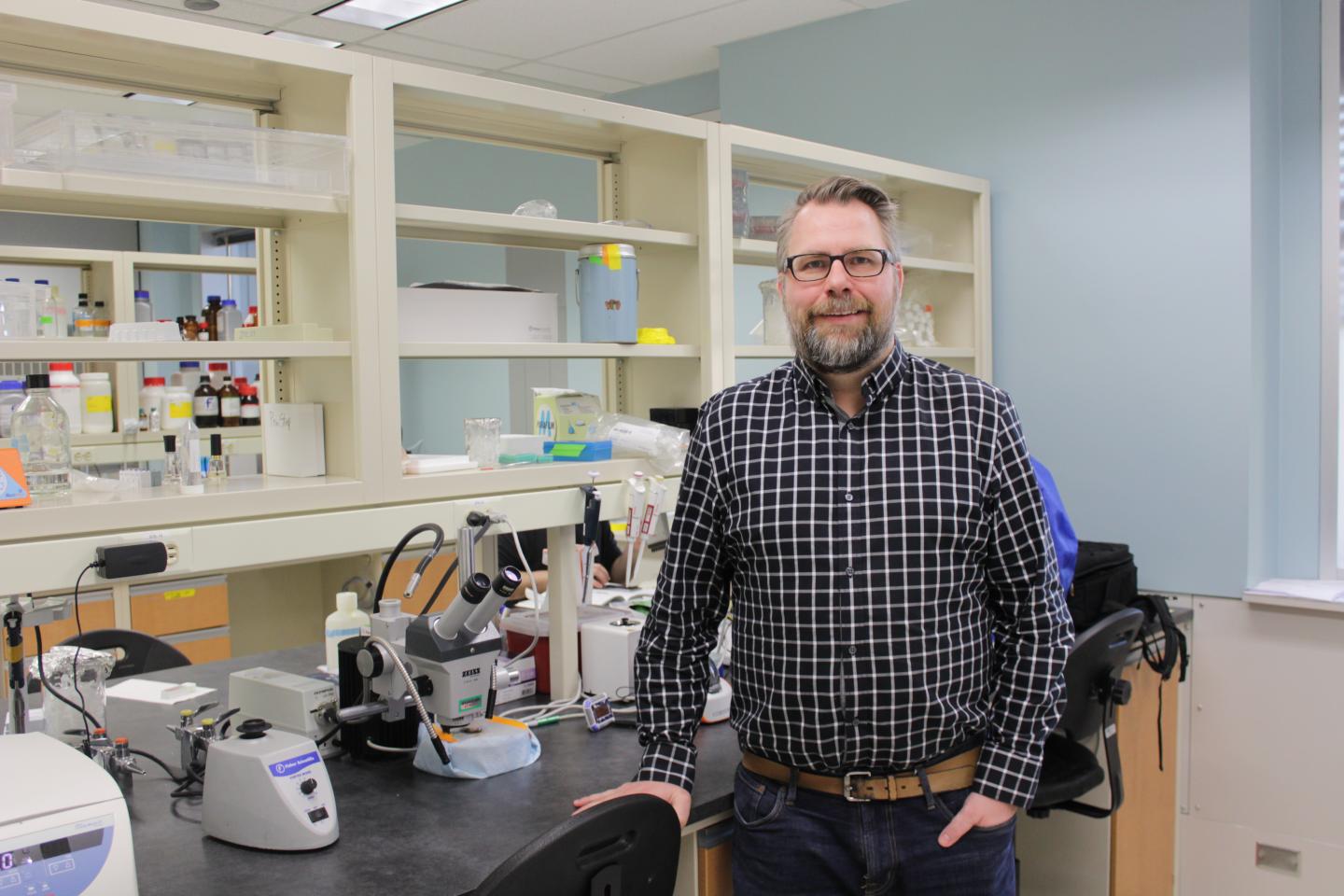
"We expected the macrophages would be present in the area of injury, but what surprised us was that microglia actually encapsulated those macrophages and surrounded them -- almost like police at a riot. It seemed like the microglia were preventing them from dispersing into areas they shouldn't be," said Jason Plemel, PhD, medical researcher from the University of Alberta, in a statement.

With the ability to differentiate between microglia and CNS-infiltrating macrophages, the researchers then performed single-cell transcriptomic analysis to understand how microglia are activated in demyelinated spinal sections. They used fluorescence-activated cell sorted (FACS)-purified tdTom+ microglia after lesion induction and prepared single-cell RNA sequencing (RNAseq) libraries from control and CX3CR1+/tdTom+ samples. A machine learning computational time-based approach showed that activation occurs in several states. Moreover, the analysis revealed increased expression of Ptprc -- the gene for CD45 -- and decreased expression of homeostatic genes, such as Siglech, Sall1, and Tmem119.
"The indication of at least two different populations of microglia is an exciting confirmation for us," Plemel said. "We are continuing to study these populations and hopefully, in time, we can learn what makes them unique in terms of function."
Immunohistochemistry analysis of the reporter (tdTom, microglia) versus biomarker (CD45) revealed expansion of microglia and reduction of infiltrating macrophages seven days after injury. Microglia became activated, then surrounded and confined the infiltrating macrophages. In microglia-negative mice, CNS-infiltrating macrophages dispersed, suggesting that activated microglia interfere with peripheral macrophage dynamics, indirectly regulating their spread.
Overall, this research shows that microglia preferentially accrue and monopolize the CNS lesion site after injury, forming several activation sites. The activated microglia expand in the CNS and confine CNS-infiltrating macrophages. This research improves the mechanistic understanding of how the body responds to myelin damage or deterioration that occurs in some neurological disorders and diseases. It may also help researchers develop more effective therapies to treat those conditions.
Do you have a unique perspective on your research related to immunology or bioinformatics? Contact the editor today to learn more.
Copyright © 2020 scienceboard.net


