


July 9, 2021 -- Researchers have captured six new structures of the ribosome and its directional movement using single-molecule fluorescence resonance energy transfer (smFRET) and cryogenic electron microscopy (cryo-EM). The results were published in Nature on July 7.
The ribosome is a key machine that powers the central dogma of biology -- that DNA is first transcribed into RNA and RNA is then translated into proteins. As it carries out its job, the ribosome undergoes many conformational changes in order to accommodate diverse mRNA and transfer RNA (tRNA) cargos. While it is known that the ribosome goes through a series of stages of engagement with mRNA and tRNA, the precise molecular and directional basis of the movements are not completely understood.
"A fundamental question in science is how molecules harness thermal and chemical energy to perform their diverse functions," said corresponding author Scott Blanchard, PhD, a principal investigator and leader of the Center for Single-Molecule Imaging at St. Jude, in a statement. "One of the real mysteries of life is figuring out how molecules navigate between various conformations, in an ordered way, to generate directional motion and work."
The St. Jude team used smFRET to guide the capture of six cryo-EM structures of the ribosome in the early and late stages of translocation. This process advances mRNA-tRNA moiety to allow the next codon to move into the decoding center.
"Our work integrates single-molecule data where we obtain molecular movement information in real time, with cryo-EM, which is a static structural method," Blanchard said. "smFRET is extraordinarily powerful in its ability to reveal an ordered sequence of events with molecular interpretations."
The work reveals for the first time how the elongation factor G (EF-G) engages with the ribosome complex.
"One thing that smFRET makes evident is that you need to add the dimensions of time and change to structural biology to really understand molecular functions and drug actions," said first author Emily Rundlet, a doctoral candidate in the department of structural biology at St. Jude.
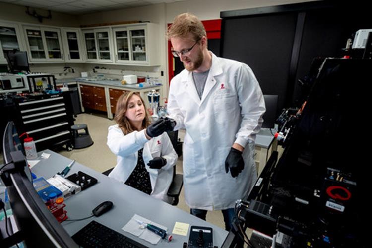
Co-authors Mikael Holm and Emily Rundlet of St. Jude Children's Research Hospital at work. Image courtesy of St. Jude.
Understanding how antibiotics interact with ribosomes
Antibiotics used to treat bacterial infections often target the ribosome. For instance, the antibiotics spectinomycin (SPC) and fusidic acid specifically stall transitions after EF-G binding. The researchers found that the antibiotic SPC interacted with the small ribosomal subunit (SSU) site of the ribosome to prevent swivel during the early stages of translocation. Fusidic acid interaction with the intermediate states 2 (INT2) structure caused a 15° rotation of the EF-G to effectively stall the translocation process.
"These findings provide a background for studying how antibiotics work, at a molecular scale, how they actually inhibit the functions of this ribosomal machine in a precise way, in a way that mediates clinical treatment of infectious disease," said author Mikael Holm, PhD, a scientist at the St. Jude department of structural biology.
"One of the take-home lessons from our work is that context is everything," Rundlet said. "Molecules are dynamic; you need to look at the actual transition state that the drug actually prevents or that the drug stabilizes to understand the full architecture of the drug-binding site, which can inform medicinal chemistry efforts to make the drugs more potent."
Do you have a unique perspective on your research related to molecular biology? Contact the editor today to learn more.
---
Copyright © 2021 scienceboard.net
Member Rewards
Earn points for contributing to market research. Redeem your points for merchandise, travel, or even to help your favorite charity.

Research Topics
Interact with an engaged, global community of your peers who come together to discuss their work and opportunities.
Conferences
Connect


