


February 18, 2020 -- A new technique using organoids, self-organizing 3D tissue models, can decipher how individual cancer cells communicate with each other and the unique signals they create. This research, published in Nature Methods on February 17, could be used to help develop personalized cancer treatments in the future.
Organoids can be used to model cell-to-cell interactions in vitro, and they represent tissues more accurately than 2D models. This makes them ideal for studying multicellular diseases such as cancer.
Post-translational modifications (PTM) -- reversible, covalent additions of small chemical entities such as phosphate-, acyl-, alkyl-, and glycosyl-groups onto modifiable amino acids -- are necessary for cellular signaling. Errors in PTM and dysregulated signaling networks are hallmarks of many diseases.
Despite the importance of PTM in disease, and the development of organoids to study diseases, the technology to analyze cell-type-specific PTM networks in organoids has not been previously developed. Neither organoids nor single-cell RNA sequencing on their own can be used to measure independent PTM networks or determine cell-type. Mass cytometry (where metal-conjugated antibodies label cellular proteins), however, can be adapted to detect PTM in heterocellular systems, like organoids.
So, researchers from University College London (UCL) created a custom multivariate-barcoded mass cytometry method to analyze single-cell PTM signaling in organoids. Using this model, they found that cell-type-specific signaling networks are intimately linked with cell state. For example, in colorectal cancer organoids, epithelial oncogenic mutations mimic signaling networks normally induced by connective tissue cells (stromal cells).
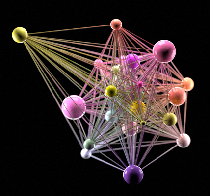
UCL artwork generated directly from data using the new method described in the paper. Images highlight signaling networks within organoid cells and signaling relationships. Images courtesy of UCL and produced by Phospho Biomedical Animation.
"Organoids are already revolutionizing cancer research by allowing us to test whether experimental new drugs are effective on lifelike models of tumors," said lead study researcher Chris Tape, PhD, from UCL, in a statement by Cancer Research UK. "But crucially, this new technique helps scientists to understand why a treatment works or not, by revealing in unprecedented detail how cells are talking to each other."
To directly compare PTM networks between healthy and diseases tissue, the researchers performed mass cytometry barcoding using thiol-reactive probes that bind to organoids in situ. They were able to simultaneously detect 28 key signaling molecules across six different cell-types in over 1 million cells. They tracked over 40 types of PTM to build detailed "circuits" that described how the cancer was working.
In the cancer organoids, the cancer cells themselves (as well as the immune cells and stromal tissues) had "rewired" normal signaling, which allowed tumors to grow unchecked. In normal tissue organoids, the cells regulated only acute signaling fluxes. The team found that colorectal cancer developed through successive oncogenic mutations where stromal cells hyper-activated the PI3K signaling pathway in colonic epithelial cells that already carried Kras and Trp53 mutations.
"Our new technology allows us to simultaneously measure the behavior of cancer cells, healthy cells, and immune cells from minitumors [organoids]," said Tape. "In healthy tissues, signals from the environment are tightly controlled so the tissue doesn't grow too fast. Unfortunately in cancer, mutations that mimic microenvironment signals are constantly switched on -- allowing the cancer to grow unchecked. The new technology developed at UCL enabled scientists to observe this phenomenon in minute detail."
Moving forward, UCL scientists plan to use this technology to study how tumors from individual patients can uniquely communicate with healthy cells and the immune system.
"By understanding how minitumors function at the single-cell level, this new technology will enable researchers to identify new ways to treat an individual's cancer," said Tape. "We expect that in the future an individual patient will have minitumors grown as 'living biopsies' alongside their clinical treatment. We will be able to test drugs on the minitumors and use that to inform how the patient's individual tumor should be treated. Unlike old approaches, our new method can also be used to study immunotherapies -- not just chemotherapies -- because it can measure lots of different cell types at once. Old methods could really only look at one cell type at a time."
Do you have a unique perspective on your research related to cell biology or cancer genomics? Contact the editor today to learn more.
---
Copyright © 2020 scienceboard.net
Member Rewards
Earn points for contributing to market research. Redeem your points for merchandise, travel, or even to help your favorite charity.

Research Topics
Interact with an engaged, global community of your peers who come together to discuss their work and opportunities.
Conferences
Connect


