


February 14, 2020 -- For the first time, scientists have made human organs transparent with the use of a new imaging technique called SHANEL, which stands for small-micelle-mediated human organ efficient clearing and labeling. The details were published in Cell on February 13.
Tissue engineering requires the use of 3D bioprinting to create cellular maps of human organs that replicate large-scale human tissues and organs. This process is limited by the complex anatomic structure and lack of scalable tools to image human tissues at the cellular level.
Magnetic resonance imaging (MRI) can provide longitudinal imaging of human organs, but it lacks resolution at the cellular level. Therefore, histology has become the primary approach to studying human organs at the molecular and cellular level. But histology has limitations of its own when it comes to 3D printing, as it involves piecing together thousands of thin slices of tissue which can be riddled with distortions caused by mechanical slicing.
Also, the thickness and complexity of human tissue make it difficult to image due to a light-scattering phenomenon that limits light penetration. The mix of components with differing abilities to scatter light (refractive index) makes tissue nontransparent. New tissue clearing technologies seek to make tissues transparent through a series of chemical reactions that try to homogenize the refractive index of all components in the sample. Despite advancements in these techniques, researchers have been unable to make human organs fully transparent.
Now, researchers from Helmholtz Zentrum München, Ludwig Maximilians University Munich, and Technical University of Munich have developed a new technique called SHANEL to address the challenges associated with making human organs transparent for imaging.
"We had to change our approach completely and start from scratch to find new chemicals which can make human organs transparent," said lead study author Shan Zhao, a doctoral student at Helmholtz Zentrum München, in a statement.
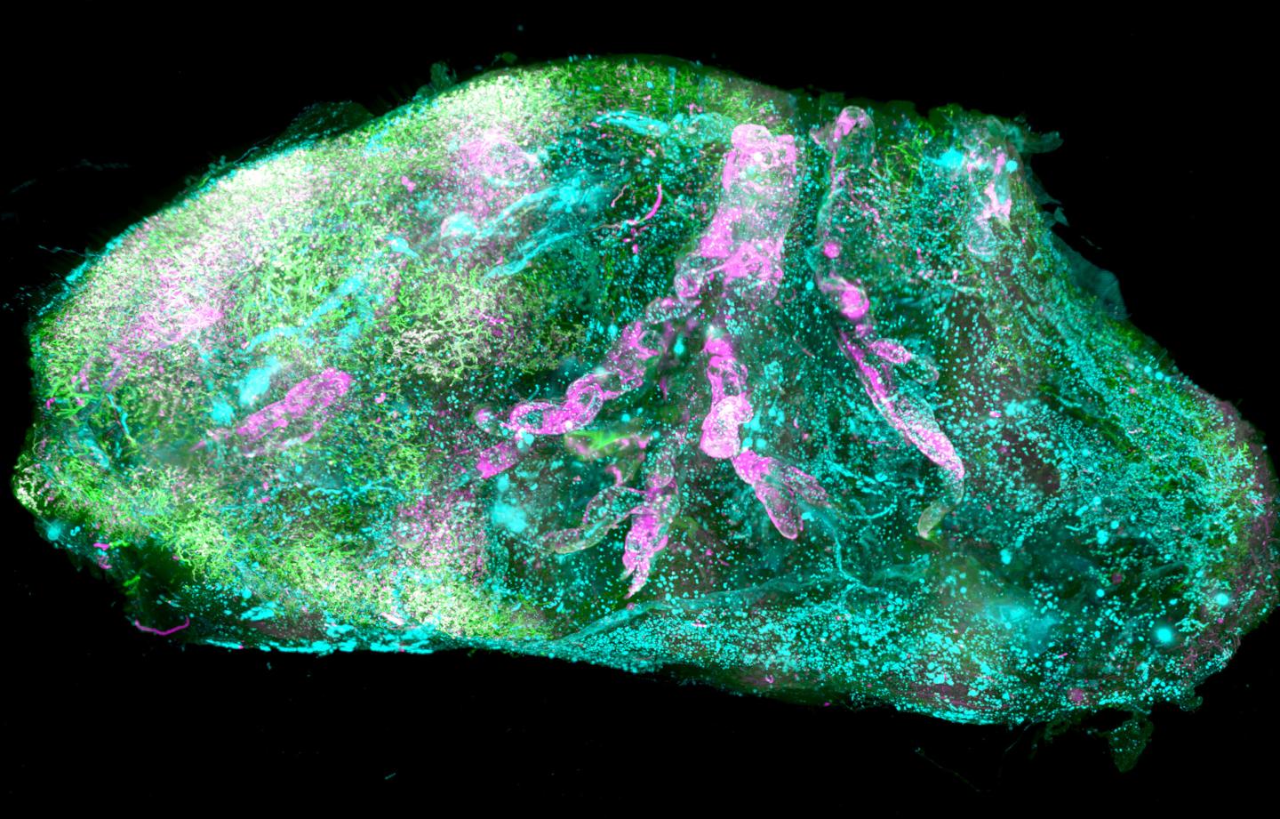
Initially, the researchers found that traditional detergents used for tissue clearing do not work on human organs. But after many tests, they found that 3-[(3-cholamidopropyl) dimethylammonio]-1-propanesulfonate (CHAPS) permeates human tissues more fully than other detergents. They used this detergent in combination with organic solvent-based clearing methods to make tissues transparent with the SHANEL technique
After making the tissues transparent, the researchers turned to imaging techniques. They developed a prototype light-sheet microscope called "Ultramicriscope Blaze" with extended stage movements and a large sample chamber. In combination, these tools allowed the researchers to study pathologies of intact human organs at a gross scale to provide new information and understanding of organ function, health, and disease.
Condensed light traveling end-to-end in a transparent human brain made visible with SHANEL. Image courtesy of Helmholtz Zentrum München and Ertürk lab.
One major issue with light-sheet microscopy is the amount of data generated from cleared tissues. Several software packages are available to detect objects and cells from the data, but recent deep-learning techniques have proven superior to the previous filter and threshold approaches. So the researchers adopted a deep-learning approach based on convolutional neural networks to analyze the data from cleared tissues. This approach was 10 to 20 times faster than previous analyses.
"SHANEL can develop into a key technology for mapping intact human organs in the near future. This would dramatically accelerate our understanding of organs such as the brain, their development and function in health and disease," explained Dr. Ali Ertürk, director of the Institute for Tissue Engineering and Regenerative Medicine at Helmholtz Zentrum München, in a statement.
In the future, the cellular maps produced by SHANEL could be used to engineer scale human tissues and organs with 3D printing technologies. The researchers are already working on mapping major human organs, including the pancreas, heart, and kidney.
"There is a huge shortage of organ donors for hundreds of thousands of people," Ertürk said. "The waiting time for patients and the transplantation costs are a real burden. Detailed knowledge about the cellular structure of human organs brings us an important step closer to creating functional organs artificially on demand."
Do you have a unique perspective on your research related to bioimaging? Contact the editor today to learn more.
---
Copyright © 2020 scienceboard.net
Member Rewards
Earn points for contributing to market research. Redeem your points for merchandise, travel, or even to help your favorite charity.

Research Topics
Interact with an engaged, global community of your peers who come together to discuss their work and opportunities.
Conferences
Connect


