


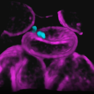
December 9, 2022 -- A new microscopy technique enhances biomedical research by capturing dynamic 3D images across larger areas while maintaining cellular resolution in all three dimensions. A study, published on December 8 in the journal Optica, provides new views of cells interacting in their natural state, potentially facilitating the development of new disease treatments.
Light diffraction limitations make 3D imaging with cellular resolution difficult over large areas. While modern photography uses additional cameras to overcome the challenge of acquiring both large scene coverage and detailed resolution, this technique isn't feasible with microscopy.
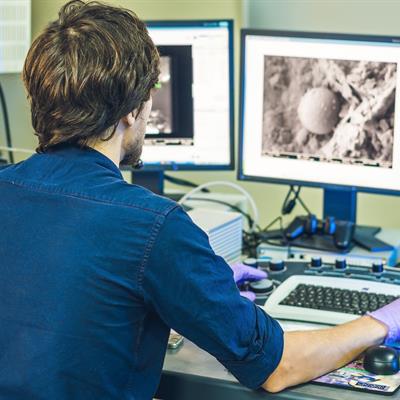
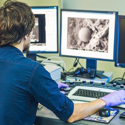
Researchers at Johns Hopkins University in Baltimore used a new mesoscopic oblique plane microscopy method, a type of light-sheet microscopy that uses a sheet of laser light to illuminate a thin sample slice labeled with fluorescent markers. Their technique captures up to three times more resolvable image points within an imaged 3D volume than similar systems.
Rather than add another camera, they used an optical component called a transmission grating to create a diffractive light sheet, which improves depth sectioning and resolution while using a low magnification lens. The result was the novel ability to perform mesoscopic scale imaging over a field of view several millimeters wide while still resolving individual cells in 3D.
The team imaged two important large-scale models: entire living zebrafish larvae 3 mm to 4 mm long and mouse brain slices containing living cells. Both expressed fluorescence proteins that labeled specific cell types, such as neurons and blood cells.
The researchers acquired whole-body volumetric recordings of zebrafish embryo blood flow dynamics at 5 Hz with 3D cellular resolution, allowing previously unseen single-cell tracking within the complete 3D circulation system. The researchers hope to improve light collection efficiency to further increase imaging speed and to incorporate multiphoton imaging to allow better penetration depth.
"Looking at biological systems in their larger context -- a mesoscopic scale -- provides holistic information for complex biological systems," Ji Yi, PhD, a co-author of the study, said in a statement. "Our work also lays a foundation for further development that would allow even faster, larger, and deeper biological imaging."


Copyright © 2022 scienceboard.net
Member Rewards
Earn points for contributing to market research. Redeem your points for merchandise, travel, or even to help your favorite charity.

Research Topics
Interact with an engaged, global community of your peers who come together to discuss their work and opportunities.
Conferences
Connect


