


July 9, 2021 -- Researchers have successfully combined multiple imaging modalities on the same brain tissue to capture the anatomic features of neurons across the entire mouse brain. The pipeline of imaging modalities and results are detailed in NeuroImage.
Magnetic resonance imaging (MRI) allows mapping of neuronal tracts spanning macroscopic distances, but cannot easily observe individual neurons, much less identify individual neuronal connections. On the other end of the spectrum, automated electron microscopy (EM) provides synapse-level resolution.
Synchrotron-based x-ray tomography (µCT) nondestructively provides micrometer-scale resolution in whole tissues and has the potential to bridge the resolution disparity between MRI and EM imaging techniques.
Furthermore, sample preparation for µCT is compatible with postmortem MRI and large volume EM. Thus, the researchers at the University of Chicago (UChicago) and the U.S. Department of Energy's (DOE) Argonne National Laboratory sought to confirm that MRI followed by µCT and EM provides a viable pipeline for multiscale whole brain imaging.
"Our lab is really interested in mapping brains at multiple scales to get an unbiased description of what brains look like," said senior author Narayanan Kasthuri, PhD, assistant professor at UChicago and a scientist at Argonne, in a statement. "When I joined the faculty here, one of the first things I learned was that Argonne has this extremely powerful X-ray microscope, and that it hadn't been used for brain mapping yet, so we decided to try it out."
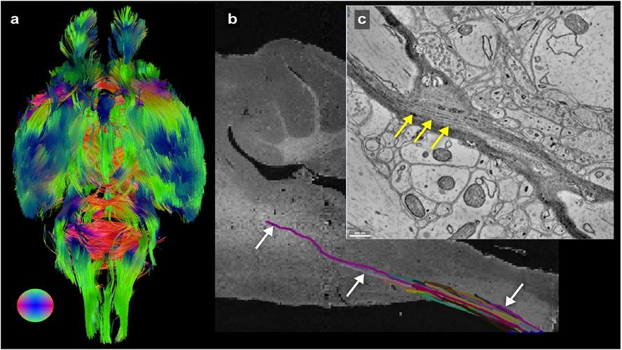
A team of scientists with Argonne and UChicago visualized an entire mouse brain from synapses on up. Above, a visualization of whole brain interconnectivity, with colors depicting directionality. Image courtesy of Sean Foxley.
Using the imaging pipeline -- MRI, µCT, and EM -- the team was able to image the same mouse brain tissue over five orders of spatial resolution (from millimeters to nanometers). The scientists demonstrated that imaging with µCT provides sufficient information and volume to help register an EM dataset to the relative low-resolution framework of an MRI volume.
The technologies enabled the researchers to track individual myelinated axons and their processes on a brain-wide level as well as identify individual synapses on the axons. The pipeline provided an unprecedented look across a single brain's multiscaled organization and will be a vehicle for studying the brain's multiscale pathologies in the future, according to the authors.
"There have been a lot of imaging studies using MRI to look at the whole brain level and then try to validate those results using electron microscopy," said first author Sean Foxley, PhD, assistant professor at UChicago. "It's hard to say anything about the large volume of tissue you see with an MRI when you're looking at an electron microscopy dataset, and the X-ray can bridge that gap."
Do you have a unique perspective on your research related to bioimaging? Contact the editor today to learn more.
---


Copyright © 2021 scienceboard.net
Member Rewards
Earn points for contributing to market research. Redeem your points for merchandise, travel, or even to help your favorite charity.

Research Topics
Interact with an engaged, global community of your peers who come together to discuss their work and opportunities.
Conferences
Connect


