


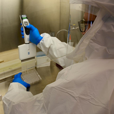
September 11, 2020 -- New images of respiratory tract cultures of the infectious form of SARS-CoV-2 produced in epithelial cells clearly show a vast amount of virus present. The images were captured by Camille Ehre, PhD, assistant professor of pediatrics at the University of North Carolina (UNC) School of Medicine.
Human bronchial epithelial cells were inoculated with SARS-CoV-2 virus in a biosafety level 3 research laboratory. The images were then captured using scanning electron microscopy and recolorized by medical student Cameron Morrison to show how the virus gets trapped in mucous that is transported to airway epithelial cells by cilia.
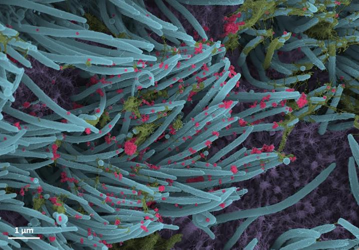
SARS-CoV-2 virions (red). Images courtesy of Ehre Lab, UNC School of Medicine.
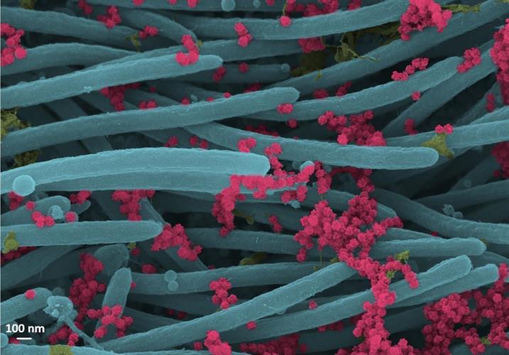
A higher-power magnification image shows the structure and density of SARS-CoV-2 virions (red) produced by human airway epithelia.
Virus production was approximately 3,000,000 plaque-forming units per culture, a finding that is consistent with a high number of virions produced and released per cell. The presence and density of virons present per cells within the respiratory tract indicates the large viral burden associated with COVID-19 and may explain the high frequency of viral transmission.
Copyright © 2020 scienceboard.net
Member Rewards
Earn points for contributing to market research. Redeem your points for merchandise, travel, or even to help your favorite charity.

Research Topics
Interact with an engaged, global community of your peers who come together to discuss their work and opportunities.
Conferences
Connect


