
April 30, 2020 -- A research project led by chemists at the University of Warwick has revealed how ultrahigh-resolution scanning tunneling microscopy (STM) can be used to determine precise molecular interactions, which could be particularly useful for the production of raw materials. The report was published in Nature Communications on April 30.
The engineering of functional molecular nanostructures for pharmaceutical use requires self-assembly via strong, directional intramolecular forces such as hydrogen or halogen binding. 2D assemblies are often characterized on surfaces with scanning probe microscopy techniques such as STM and atomic force microscopy.
Recently, functionalized tips (such as CO, Xe, D2, H2, Br, and CuO) have been used to achieve higher resolution of internal structures. STM has been used to resolve structures that are difficult to determine with more traditional analytical methods such as nuclear magnetic resonance or mass spectrometry.
The researchers used ultrahigh-resolution STM to see the exact location of atoms and bonds within the self-assembly of 3,9-dibromo-peri-xanthenoxanthene (3,9-Br2PXX) molecules on a gold surface. They used a carbon monoxide tipped super sharp needle frozen to 7 degrees Kelvin (-447 degrees Fahrenheit) to clarify the structure of the bromide molecule. The results indicated that the structures were governed by halogen-bonding interactions.
Next, the researchers compared standard and ultrahigh STM, using density functional theory calculations as a map of the electronic structure of the bromide molecule. They demonstrated that standard STM could not conclusively establish the nature of the intermolecular interactions, but the new technique could clearly identify the location of carbon rings and halogen atoms.
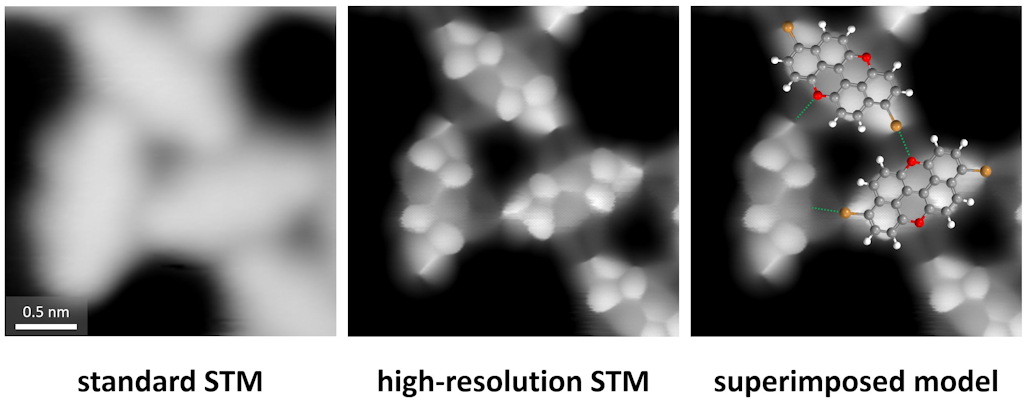
Microscope images of the bonds progressing in clarity. Image courtesy of the University of Warwick.
"Scanning tunneling microscopy (STM) can normally only reveal the overall shape and position of molecules in a material but hasn't got the precision needed to determine their exact atomic structure," said senior author Giovanni Costantini, PhD, professor in the department of chemistry at the University of Warwick, in a statement. "However, using ultrahigh-resolution STM, we could precisely pinpoint the location of carbon rings and halogen atoms, which allowed us to establish that halogen rather than hydrogen bonding governed the molecular assembly of this material."
Using the same ultrahigh-resolution STM methods, the researchers were able to detect minute impurities in the sample that other conventional spectroscopic characterization techniques fail to identify. This provides evidence that submolecular resolution scanning probe techniques such as ultrahigh-resolution STM have the potential to be powerful analytical tools for identifying the chemical structure of unknown molecules or for determining the intermediates and products on of on-surface reactions. According to the authors of the study, the technique could provide analytical insight into the nanoscale arrangement of molecules within a supramolecular structure and the nature of the intermolecular interactions ruling the self-assembly.
"The ability to discern and actually clearly identify the position of halogen bonds will be of particular value to researchers attempting to understand biomolecular recognition and design novel pharmaceuticals drugs," explained co-author Gabriele Sosso, PhD, associate professor in the department of chemistry at the University of Warwick. "In fact, most of the medicinal chemistry up to now has been focusing on the role of hydrogen bonds, as they are ubiquitous in both biochemistry and materials science: Understanding halogen bonding will thus provide an additional tool to engineer the next generation of molecular systems for drug design. To that end, it is essential that, as we did in this work, we bring together experiments and simulations -- in order to deliver a comprehensive picture of this still largely unexplored molecular interaction."
Do you have a unique perspective on your research related to bioimaging or microscopy? Contact the editor today to learn more.
---
Copyright © 2020 scienceboard.net
Member Rewards
Earn points for contributing to market research. Redeem your points for merchandise, travel, or even to help your favorite charity.

Research Topics
Interact with an engaged, global community of your peers who come together to discuss their work and opportunities.
Connect


